Ct Scan Orbital Floor Mesh
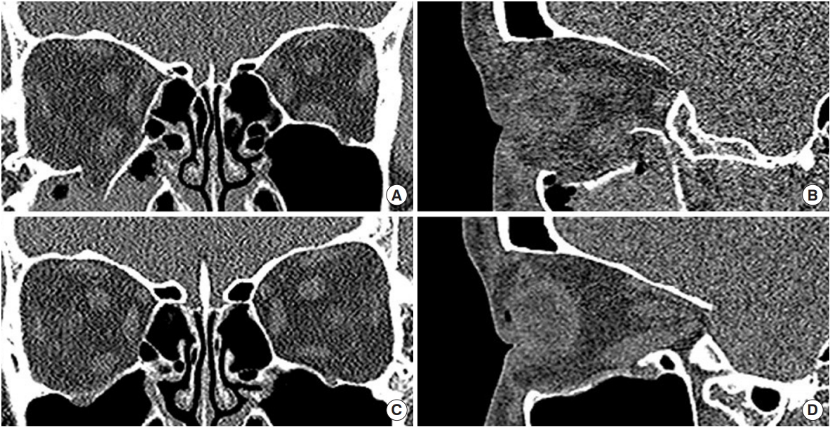
Before the advent of high resolution ct several articles were published in the radiology literature debating the need for direct sagittal views which required cumbersome patient positioning.
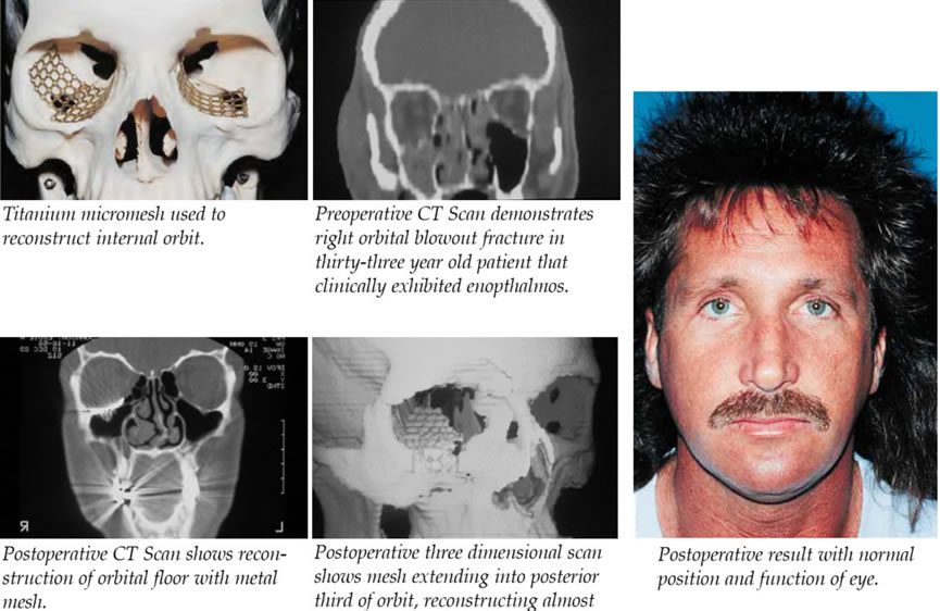
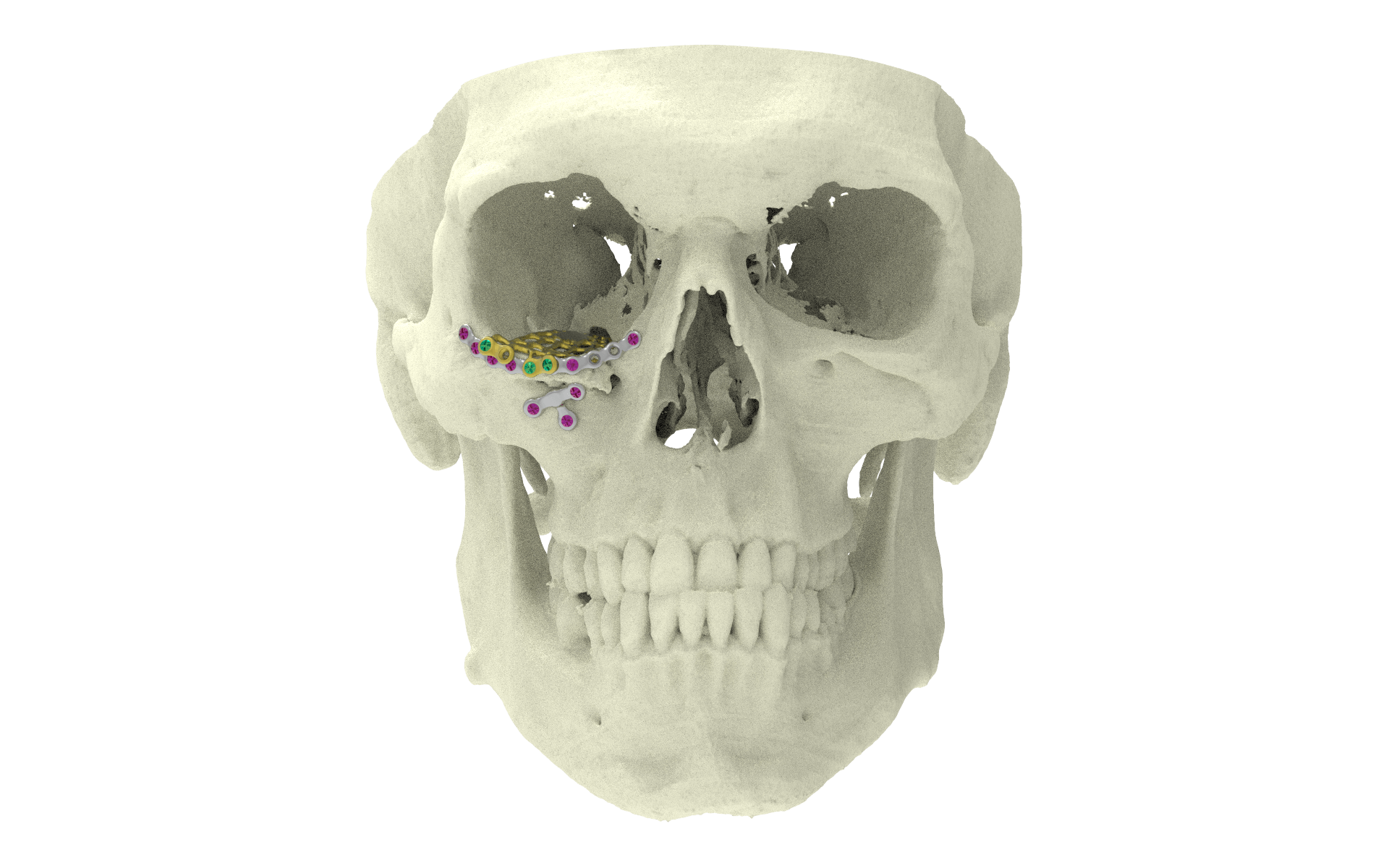
Ct scan orbital floor mesh. The matrixmidface preformed orbital plates are designed from ct scan data. In intervention group n 5 the ct scan slices were used for generating 3d reconstruction of both affected and unaffected orbits. For most orbital fractures the imaging study of choice is ct scan. Orbital fractures pose specific challenge in its surgical management.
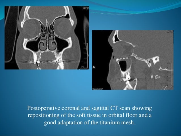
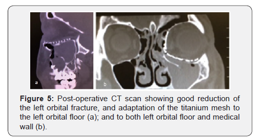
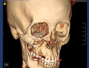
Surgical treatment was performed using subciliary inferior palpebral approach to explore the orbital floor and placement of the titanium mesh and an intraoral antrostomy for endoscopy to magnification of the surgical field and adaptation of the mesh. The aim of this study was to describe t. This confirms that there is no need for further corrections in this case. For minimal bending and cutting which reduces the amount of time.
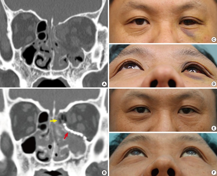
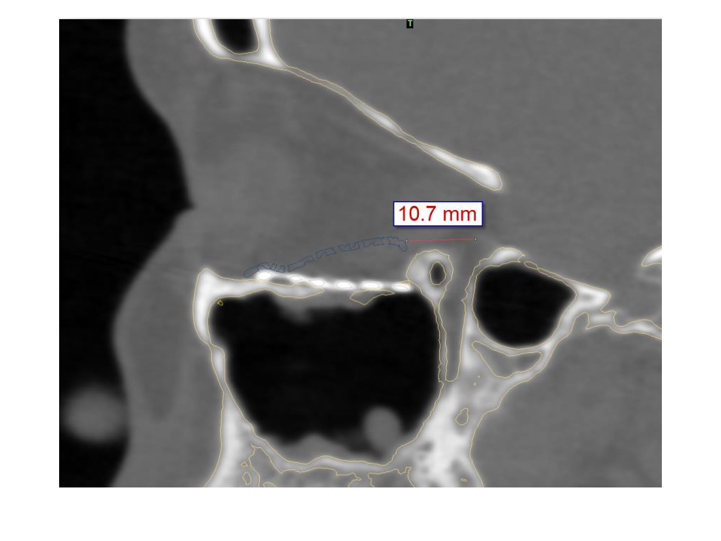
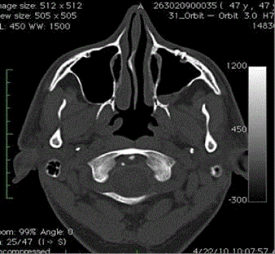
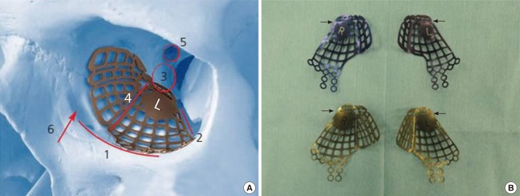
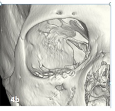
Psi placement over failed pre bend mesh. To design implants for orbital reconstruction rapid prototype models can be derived from digital imaging and communications in medicine dicom data obtained from the patient s computed tomography ct scan. The overlying colored line in the medial wall and orbital floor area indicate the preoperative virtual planning that is superimposed on the mesh reconstructed area. A ct scan with axial and coronal views is optimal.
One of the greatest challenges is to obtain satisfactory reconstruction by correct positioning of orbital implant. Orbital floor designed from ct scan data the three dimensional implants closely approximate the topographical anatomy of the hu man orbital floor and medial wall to provide accurate recon struction even after significant two wall fractures 5 6 preformed three dimensional shape. This study was prospectively conducted on 10 patients with unilateral orbital floor fractures caused by accident or falls. These plates consist of implants that closely approximate the topographical anatomy of the human orbital floor and medial wall and are intended for use in a selective craniomaxillofacial trauma.
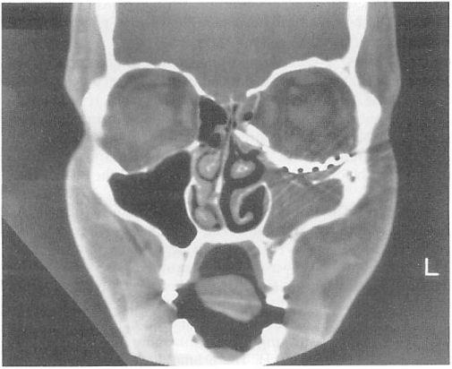
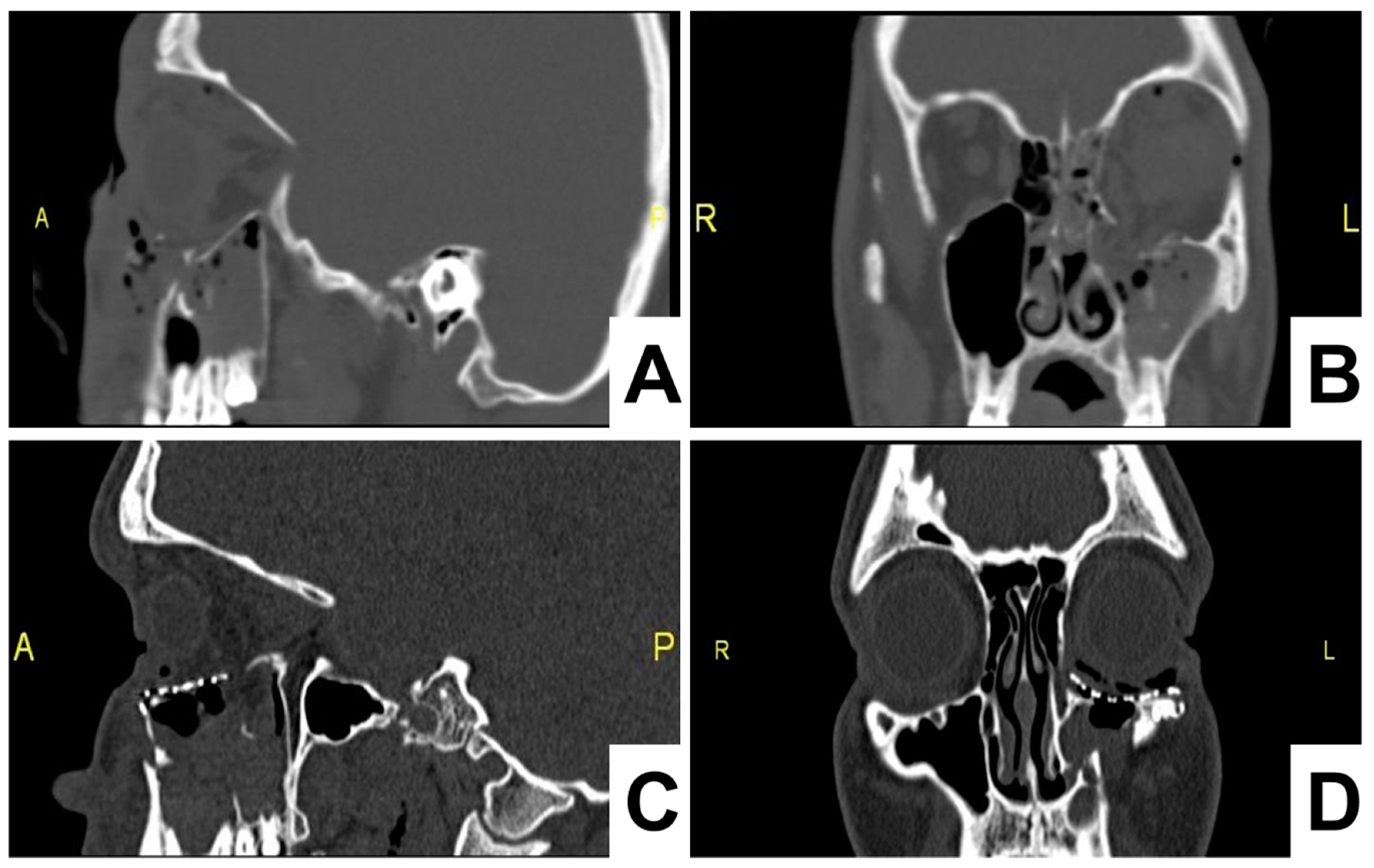
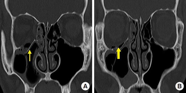
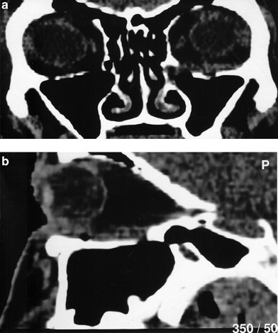
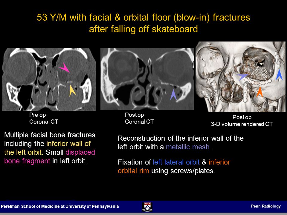
The sagittal plane computed tomographic ct scan has been proposed as the most important radiologic view in the diagnosis of orbital floor fractures. Postoperative ct scan analysis shows that all treatments restored orbital volume and. Coronal slice of a postoperative ct scan taken after transconjunctival repair of the complete left medial orbital wall and orbital floor. This x ray shows the classic transition zone.
In case the orbital floor is not properly reconstructed correction of shape and position of the implant is recommended followed by a. Ask for thin cuts 2 3 mm with specific attention to the orbital floor and optic canal.


